
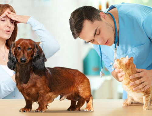
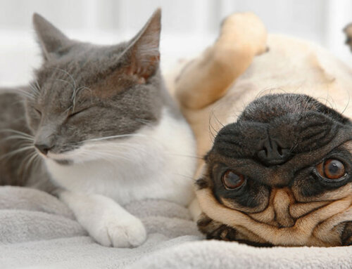
I was talking with a kitty owner today. Her kitty stopped eating earlier this week. We ran bloodwork and took radiographs and then sent her to an internist for an ultrasound yesterday. We still don’t have an answer as to why her kitty isn’t eating, but we’re treating with antibiotics, anti-nausea meds and an appetite stimulant. There were some suspicious abnormalities on Radiographs and the ultrasound revealed some thickening of the stomach and small intestine. The internist may repeat the ultrasound or I may repeat X-rays if our sweetie doesn’t turn a corner soon. As we were discussing our plan of action I thought how this would be a great topic for an ADW newsletter.
My mission at ADW Diabetes is educate our readers about diabetes. A lot of our newsletters concentrate on glucose testing and glucose curves and diet, but if a pet’s diabetes isn’t regulating we may do diagnostics to look for an underlying issue. Imaging is a common diagnostic tool, so today we will discuss types of imaging and when we might choose one over another.
Radiographs
Radiographs are commonly called X-rays. Radiographs are 2 dimensional snapshot images. They are a moment in time. We rarely take a single image of a body part. Routinely we will take several views so that we can imagine what is happening from these 2D images. In the age of digital radiology (which gives us images much faster than the old fashioned films which needed to be processed), I now routinely take 3 views of a chest or abdomen. Radiographs are essentially shadows. They show us gas or fluid or mineral densities. Now if we want more than just a moment in time, there is fluoroscopy which is real-time X-ray. Very few vet clinics have a fluoroscope. Fluoroscopy is especially great for diagnosing airway issues, but fluoroscope are mostly at veterinary teaching hospitals at veterinary schools. Most veterinary practices have radiographs.
Ultrasound
Ultrasound is bouncing sound waves back from an object and making an image based on the return of those waves. Ultrasound gives us an idea of what is within an organ, if the organ is homogeneous or if there is a mass or irregularity. Ultrasound doesn’t work particularly well if there is bone or gas in the way, but for soft tissue it can provide wonderful images, particularly with the newer machines. When a woman goes in for pregnancy imaging it is usually an ultrasound the doctors use. This is a real-time image but we can freeze the screen and save a permanent image. Or, we can make a brief video, say a five second loop. With saved images we can review the image or take measurements of an organ or mass. Or, if it is an image of your baby on the way you can show the image to your family and friends!
CT And MRI
CT and MRI both provide images of a body part in cross section. CT is usually faster and less expensive than MRI. CT is commonly used in human emergency rooms and is particularly good for bones and lungs. MRI provides better soft tissue detail than does a CT scan. MRI is particularly good for brain and spinal cord images. These machines are pretty pricey, so the average veterinary practice won’t have one. I remember years ago the local neurologist used to do his patients’ MRIs after hours at a human facility. These days most big cities will have a veterinary specialty hospital that has either a CT or MRI machine.
Endoscopy
Endoscopy is putting a teeny little camera on a flexible tube into some body part. For example, a colonoscopy is sticking a camera up someone’s bum. Rhinoscopy is sticking a camera up a nose. Laparoscopy is doing this into the abdomen. Arthroscopy is putting a camera into a joint. You get the idea. It’s amazing how small these cameras can be! Endoscopy for pets usually requires anesthesia or at least heavy sedation. Most endoscopes have a separate channel where a biopsy instrument can be slid alongside the camera’s tubing so that a biopsy of the tissue can be taken. This is a minimally invasive way of both visualizing an area but also taking tissue samples. Some general practices have endoscopes, but more often than not you will be referred to a specialist of your pet needs endoscopy.
You know I like hearing from our readers. Don’t hesitate to email me at [email protected]
NOTE: Consult your veterinarian to confirm that my recommendations are applicable for the health needs of your pet.


Leave A Comment